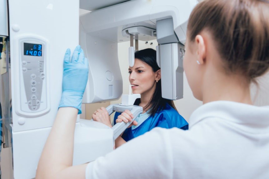
When it comes to imaging technology for dental patients, there are a number of choices. Both OPG and CBCT offer excellent resolution, clarity, and precision. They also both provide a 3D image that aids in the diagnosis of potential problems. But how do these two technologies differ? Both intraoral photography and cone-beam computed tomography (CBCT) share the same basic principle: imaging many slices of the oral structures to produce a detailed, three-dimensional picture of the patient’s teeth, bones, and soft tissues. These diagnostic tools provide different methods for increasing visual detail. For example, when compared to traditional panoramic images or standard intraoral photography, CBCT reveals greater detail about the shape and structure of soft tissue — including minor irregularities invisible to the human eye. Likewise, OPG offers visibility into smaller crevice angles than standard radiography or contact lens cameras while still producing an image comparable in quality to CBCT.
OPG or Orthopantomogram
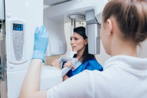
OPG is an acronym that stands for “oral panoramic.” OPG is a dental x-ray device in which the patient bites down on an acrylic dental bite registration device while a technician takes an image of the teeth with a low-dose x-ray. A OPG is often used to take a full-mouth image of the teeth and supporting structures to identify potential dental problems that could include misaligned teeth, diseased teeth, or missing teeth.
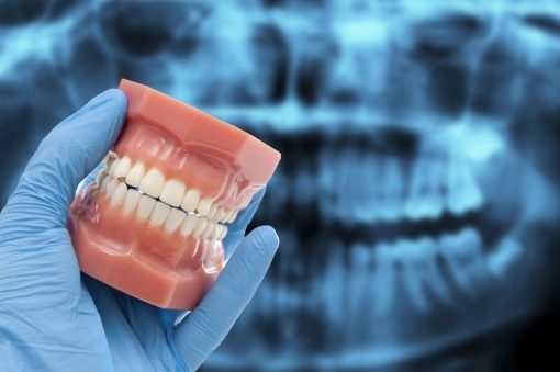
OOP images can be stitched together to create a panoramic view of the dental arches. OOP images are usually higher in resolution than CBCT images, making them more suitable for use in diagnosis. A OOP images are also more suitable for diagnosis when the patient is edentulous or does not have any teeth, as CBCT is not suitable for patients with no teeth. OOP devices are often more affordable than CBCT devices and are available in standalone devices that can be used in any dental practice.
CBCT or Cone-beam computed tomography
CBCT is an acronym that stands for “computed tomography.” CBCT is an imaging device that uses x-rays to produce a high-quality, three-dimensional image of the teeth and surrounding tissues. A 3D imaging device may also be called a CT, Cone Beam, or CT Scanner. However, the images taken with a CBCT device are different from those taken with OPG in a number of ways: 3D images produce more detailed visualisations of soft tissue than OPG.
CBCT images are stable and can be taken in any position — unlike the images from OPG, which require the patient to remain in a fixed position. CBCT images do not require tissue retraction. 3D images are more suitable for diagnosis when the patient is edentulous or does not have any teeth. These distinctions between OPG and CBCT allow dentists to select the imaging technology that is most suitable for each individual patient.
Differences in quality and image resolution
Both OPG and CBCT provide excellent image resolution. That said, CBCT images are generally higher in resolution than those taken with OPG. Depending on the type of machine and settings used. 3D images can be up to 10 times higher in resolution than those taken with OPG.
Differences in soft tissue visibility | CBCT vs OPG
Compared to OPG, CBCT imaging provides better visualization of soft tissues. While this is advantageous for general dentistry, CBCT images are not suitable for diagnosing pathologies in oral soft tissues — such as fibrous tumors, cysts, and cysts. Because of its greater soft tissue visibility, CBCT is more suitable for dentists who specialize in endodontics, periodontics, orthodontics, and oral pathology.
Differences in Stable vs Variable Imaging Positions
Its imaging requires the patient to remain in a fixed position during the imaging process. Alternatively, CBCT imaging is stable and can be performed in any position. While the patient is moving or while they are standing still. For some dental procedures, the patient’s ability to remain in the same position during imaging is crucial — especially when imaging the teeth or surrounding bones.
When this is the case, OPG is the more suitable imaging choice. The imaging position of CBCT is advantageous in certain situations, such as when imaging the palate in edentulous patients. In these cases, CBCT imaging can be performed with the patient standing, face down, sitting down. Or in any other position that is most suitable for the procedure.
Differences in Probing Ability
OPG is suitable for detecting dental caries, dental root caries, and dental periodontal disease. CBCT is suitable for diagnosing dental caries, dental root caries, dental periodontal disease, and periodontal abscesses. While both imaging procedures are suitable for detecting dental caries, 3D imaging is more suitable for detecting dental root caries. CBCT is also more suitable for diagnosing periodontal disease and periodontal abscesses. While OPG can detect periodontal disease and periodontal abscesses. CBCT is more sensitive and can detect these conditions at an earlier stage.
Conclusion | CBCT & OPG
Dentists can benefit from the use of both imaging. A imaging is a suitable choice for general dentistry and for patients who cannot remain in a stable. Static position while imaging. While CBCT imaging is more suitable for certain dental procedures. It is still a good idea to have OPG images taken when 3D imaging is unsuitable for the patient. In this case, It will be the best imaging choice for the patient since it does not require any teeth. By understanding the differences between OPG and CBCT, dental professionals can select the imaging technology best suited for each patient.

