
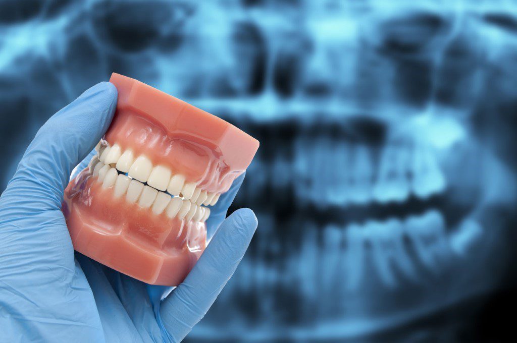
When you visit the dentist, you expect them to give you a check-up and clean your teeth. You probably don’t expect that check-up to include an X-ray. But dentists have been using X-rays as part of their check-ups for more than a century. In fact, dental X-rays are one of the oldest and most widely used diagnostic tools in modern medicine. Dental X-rays provide dentists and other healthcare professionals with valuable information about the structures inside our mouths. They can show if there are any cavities or other problems with our teeth, how tightly anchored our fillings are, whether we have any fractured or broken teeth and whether there is decay hidden behind other teeth that haven’t yet affected the surface of our mouth.
What are dental x-rays?
X-rays are a form of electromagnetic radiation. Electromagnetic radiation is made up of photons. Photons are a form of energy that travels in waves. X-rays are one type of electromagnetic radiation that can pass through most objects. We usually encounter other types of electromagnetic radiation in our daily lives, like visible light, radio waves, and microwaves.

Dentists use X-rays because they can help us see inside our mouths and diagnose problems that we can’t see otherwise. X-rays are produced by using a machine that produces a small amount of radiation. The machine uses a pulsed X-ray beam to generate an image of the inside of your mouth. Dental X-rays are not the same as the X-rays that are used to take an image of your bones. Although all X-rays cause some amount of radiation, dental X-rays are the lowest dose and lowest risk X-rays taken in any medical setting.
Why are dental x-rays used?
Dental X-rays are used for two main reasons: They allow your dentist to see inside your teeth to find problems, and they can be used to create a record of your teeth for future reference. The first reason that your dentist might choose to take an X-ray is that X-rays allow your dentist to detect cavities or other problems with your teeth that they might otherwise miss. Taking a quick X-ray can be a quick and easy way for your dentist to spot problems inside your teeth that might not be visible to you or to the naked eye.
X-rays are also a useful tool for dentists who might be considering performing a procedure or surgery on a patient. The X-ray can be used to see what is happening inside the mouth and inside of the teeth. X-rays allow dentists to see whether there are any hidden structures or cavities that could complicate the procedure. An X-ray may also be taken before a tooth extraction to make sure that the dentist removes all of the tooth fragments.
Are dental x-rays dangerous to your health?
Dental X-rays are not dangerous for the vast majority of people who receive them. The radiation from dental X-rays is less than 1% of the amount of radiation that you would receive from spending a day in the sun, and less than 5% of what you would receive from a single flight from New York to California. X-rays have been used in dentistry for many years and have proven to be a safe method of diagnosis. There is a risk, however, that overexposure to X-rays can result in health problems. This risk is much lower for dental X-rays than for diagnostic X-rays used in other fields such as radiology.
What is the danger of dental x-rays?
The danger in dental X-rays is the amount of ionizing radiation that they produce. Ionizing radiation is a known carcinogen. However, the amount of radiation that you receive from a dental X-ray is extremely small. A single dental X-ray delivers less than 0.1% of the radiation exposure that is considered to be a safe exposure to ionizing radiation. For comparison, a CT scan can deliver a dose of ionizing radiation that is 1,000 times higher than a single dental X-ray.
Dental X-rays are useful because they allow dentists to see the inside of your teeth, but they can only do that by letting a small amount of ionizing radiation pass through your teeth. This radiation is not enough to cause any serious health problems. In fact, the amount of radiation that you receive from a single dental X-ray is too small to cause any harm.
Benefits of dental x-rays for your teeth and mouth
The main benefit of dental X-rays is that they allow dentists to see inside your teeth and detect cavities or other problems before you have any symptoms. This allows them to treat any problem before it becomes a serious dental disease. Dental X-rays are also a useful diagnostic tool for dentists who are performing procedures or surgery on your teeth. X-rays allow them to see the exact location of your teeth and the position of your roots.
This allows them to more accurately plan surgery and avoid causing harm to other nearby structures. X-rays can also be used to create a record of the structure of your teeth. This can be helpful, especially if you have a condition such as periodontal disease, or have had dental work done, that causes your teeth to shift. An X-ray can show your dentist the condition of your teeth before they begin treatment or have any problems.
Should you be concerned about dental x-rays?
The main concern with dental X-rays is that they expose you to a small amount of ionizing radiation. This is a small amount of radiation compared to other common sources. You are exposed to a similar amount of ionizing radiation every time you walk outside on an overcast day. X-rays are a useful tool for dentists that provide valuable information about the health of your teeth.
However, it is also important to take steps to limit your exposure to radiation. You can lower the amount of radiation you receive from X-rays in your dental visits by keeping your appointment time as short as possible. You can also request that your dentist use a low-dose X-ray machine or take more than one X-ray view to get the same quality of the image.
Final Words – The Verdict on Dental X-ray Safety
Dental X-rays are a valuable diagnostic tool for dentists and other healthcare professionals. Although they do expose you to a small amount of ionizing radiation, the amount of radiation from a dental X-ray is too small to cause any serious health problems. Taking steps to keep your appointment time short and requesting that your dentist take more than one X-ray view can help you to minimize your exposure to X-rays. If you are concerned about the amount of radiation you are receiving, speak to your dentist about ways to lower your exposure.