Dental radiology, a crucial element in modern dentistry, is vital for diagnosing and treating various oral health issues. Renowned dentist Dr. Chirag Chamria guides us through the intricacies of this field, shedding light on its relevance, methods, and its pivotal role in ensuring our oral well-being with his deep knowledge and practical expertise.
What is Radiology in Dentistry?
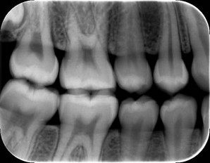
This particular kind of diagnostic imaging specializes in taking pictures of the oral and maxillofacial areas. Dental radiographs, sometimes referred to as X-rays, are important for detecting and treating a range of dental and oral health disorders. Different radiographic methods, including intraoral and extra oral X-rays, are used in dental radiography to view the teeth, jaws, and surrounding tissues.
Importance of Dental Radiology in Dentistry
Dental radiography is a crucial component of contemporary dentistry and is crucial in both the diagnosis and treatment of oral diseases.
To see the interior anatomy of the teeth, gums, and jawbone, radiographic pictures are used. Dental radiographs assist dentists in spotting dental issues such tooth decay, periodontal disease, and bone loss that may not be obvious to the naked eye.
With the advent of digital radiography, dental radiology has dramatically advanced over time. Modern radiography employs digital sensors to record radiographic pictures, which are subsequently shown on a computer screen.
This process is known as digital radiography. Dental radiology has undergone a revolution because to this technology, which produces high-quality pictures with less radiation exposure.
Types of Dental Radiology Techniques
- Intraoral Radiography:
- Purpose: Captures detailed images of individual teeth.
- Setup: X-ray film or digital sensors placed inside the mouth.
- Extraoral Radiography:
- Purpose: Examines larger areas, including jaw and skull.
- Setup: X-ray machine outside the mouth, producing panoramic or cephalometric images.
- Panoramic Radiography:
- Purpose: Captures a wide view of the entire oral and maxillofacial region.
- Setup: Patient’s head remains stationary, while the machine rotates around them.
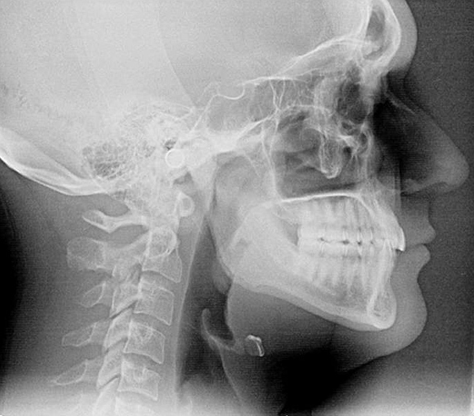
- Cephalometric Radiography:
- Purpose: Focuses on capturing side-view images of the head to assess facial structure.
- Setup: Machine positioned to take images from a specific angle.
- Cone Beam Computed Tomography (CBCT):
- Purpose: Provides detailed 3D images for complex diagnostic and treatment planning.
- Setup: Cone-shaped beam rotates around the patient, capturing images from various angles.
Advantages and Limitations of Dental Radiology
| Advantages of Dental Radiology | Limitations of Dental Radiology |
|---|---|
| Prior to being obvious to the naked eye, dental disorders including cavities, gum disease, or cysts may be identified using dental radiography. This makes it possible for prompt treatment and action. | Dental radiography uses modest quantities of radiation, yet prolonged exposure may still be dangerous. Dentists should only make sparing and required use of this instrument. |
| Detailed and reliable information about the teeth and adjacent tissues is provided by radiographs, which aid in the diagnosis. This aids in the accurate diagnosis of diseases including impacted teeth, bone anomalies, and oral infections by dentists. | Installing and maintaining dental radiography equipment, particularly digital systems, may be expensive. The fees imposed on patients could include a deduction for this expense. |
| Radiographs provide dentists the ability to keep an eye on the development of ongoing treatments and make sure they are moving along as planned. This is crucial for dental implants and orthodontic procedures. | Utilizing radiographic equipment requires implementing effective infection control procedures in order to avoid cross-contamination. |
Common uses of Dental Radiographs
They are useful for identifying cavities, evaluating the condition of the bone that supports the teeth, and identifying dental issues such abscesses, fractures, and impacted teeth. For difficult dental operations including root canals, dental implants, and orthodontic therapy, radiographs are also utilized to plan and carry out the surgery. Radiographs provide dentists an in-depth image of the mouth, enabling them to precisely plan and carry out these operations.
Safety precautions and Radiation protection
Radiation is utilized in dental radiology, which may be dangerous if improperly applied. Dentists use lead aprons and restrict the number of radiographs they take as a few of the steps they take to reduce radiation exposure.
Digital radiography, which produces high-quality pictures with less radiation exposure, is also used by dentists. Patients are safer because digital sensors need less radiation exposure than conventional film radiography.
Role of Digital Radiography in Modern Dentistry
Dental radiology has undergone a revolution because to digital radiography, which offers high-quality pictures with less radiation exposure. Patients are safer because digital sensors need less radiation exposure than conventional film radiography.
Additionally, it gives dentists access to a number of techniques and technology that enable them to precisely detect oral diseases. Dentists may save and distribute radiographic images electronically thanks to digital radiography.
With the aid of this technology, dentists may view radiographs at any time and from any location, making it simpler to work with other dental specialists on challenging situations.
Dental Radiology view from Dr. Chirag Chamria
With significant training in zygomatic implants, cosmetic dentistry, orthognathic surgery, whole-mouth rehabilitation, and oral cancer treatment, Dr. Chirag Chamria is a Certified Oral and Maxillofacial Surgeon.
The appointment of Dr. Chirag as Director and Maxillofacial Surgeon by Royal Health Care Pvt Ltd is an honor. He is presently an attending physician at Royal Dental Clinics, where he practices oncology, facial aesthetics, and cosmetic dentistry as a dental surgeon and top dentist in Mumbai.
Dental radiology, in Dr. Chamria’s opinion, is an essential component of contemporary dentistry that enables dentists to precisely detect and treat oral diseases. He stresses the value of employing digital radiography since it produces pictures of excellent quality with less radiation exposure.
Conclusion
It is clear that dental radiology is still a developing discipline. It is helping to influence contemporary dentistry as we come to a conclusion with this informative essay on it. The development of imaging technology like digital radiography and 3D imaging has completely changed how dentists identify and treat issues relating to oral health. Immerse yourself in the field of dental radiography, accept the power of the pictures, and see the revolutionary changes it has brought about in oral healthcare.

