
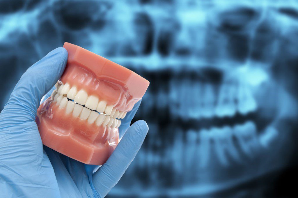
X-rays of the teeth have long been recognized in the area of dentistry as a very helpful diagnostic technique. Dentist are able to uncover previously undetected dental issues and arrive at an accurate diagnosis. However, there are worries regarding the safety of Multiple Dental X-Rays exposure. These issues have been raised in recent years. In the following paragraphs, we will investigate the question of whether or not it is risky to have multiple dental X-rays in a span of thirty days. In order to have a better understanding of the situation, we consulted the knowledge and experience of Dr. Chirag Chamria, an oral and maxillofacial surgeon who practices at Royal Dental Clinics. Repeated dental X-rays for treatment, raises safety concerns.
Understanding Dental X-ray
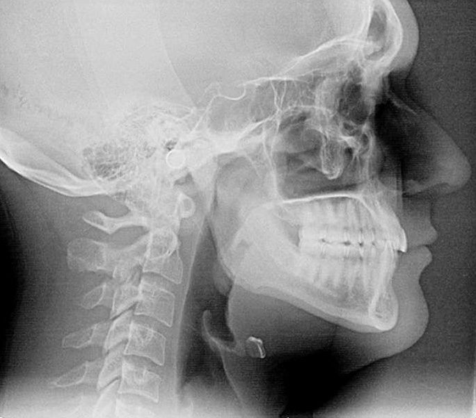
They are also known as dental radiographs, play a crucial role in modern dentistry. They are diagnostic tools used by dentists to examine and evaluate the health of a patient’s teeth, gums, and supporting structures. By capturing detailed images of the oral cavity, they enable dentists to identify dental issues that may not be visible during a regular dental examination.
Different Dental Imaging Procedures expose patients to different amounts of radiation:
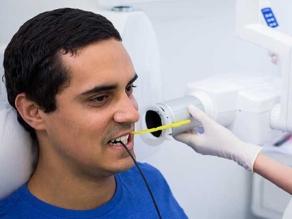
- Intraoral X-ray: Captures one or a few teeth and generally has a very low radiation dose.
- Bitewing X-ray: Helps detect cavities between teeth and evaluate the supporting bone.
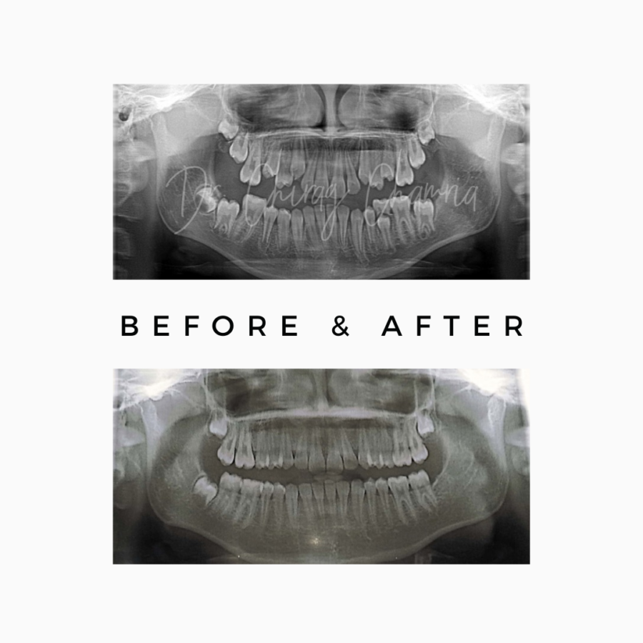
- Panoramic X-ray or OPG: Captures both jaws and a broader view of the teeth.
- CBCT scan: Produces three-dimensional images for procedures such as implant planning, evaluation of impacted teeth and assessment of complex jaw conditions.
Benefits of Dental X-rays for Dentist
- Early Detection of Dental Problems: Dental X-rays might detect hidden problems. They assist dentists discover cavities, infections, impacted teeth, cysts, tumors, and bone loss. Early identification and treatment reduce problems.
- Accurate Diagnosis: X-rays help diagnose. They help dentists diagnose and treat dental issues.
- Monitoring Dental Health: Monitoring dental procedures requires dental X-rays. They help dentists evaluate root canal and orthodontic procedures.
- Treatment Planning: X-rays help dentists plan therapy. They advise on extractions, implants, orthodontics, and oral surgery.
Radiation Exposure and Safety Measures
One concern often raised about dental X-rays is radiation exposure. X-rays do emit radiation, but the amount is minimal. Dental professionals take necessary precautions to minimize radiation risks and ensure patient safety.
- Modern X-ray Technology: X-ray technology has greatly decreased radiation. Digital X-rays provide high-quality pictures with less radiation than film-based ones.
- Lead Aprons and Thyroid Collars: Lead aprons protect X-ray patients from radiation. Radiation-sensitive thyroid collars protect it.
- Proper Technique and Equipment: Dentists use typical radiation-reducing methods. They calibrate and maintain X-ray equipment for precise imaging with less radiation.
- Individualized Approach: Age, medical history, and dental requirements determine X-ray frequency and kind. The ADA advises dentists on dental X-ray usage.
Frequency of Dental X-rays Recommended
The frequency of dental X-rays varies from patient to patient. It depends on factors such as the individual’s oral health, age, risk of dental diseases, and any specific symptoms or concerns. We consider these factors when determining the necessity and timing of X-ray examinations at Royal Dental Clinics.
New Patients: For new patients, X-rays are often taken as part of the initial examination. These X-rays provide a baseline understanding of the patient’s oral health and assist in detecting underlying issues.
Recall Examinations: Dr. Chamria may prescribe X-rays during recall exams or regular check-ups to monitor oral health, discover new issues, or evaluate prior procedures. Dental health may need X-rays every few months or years.
Specific Dental Issues: Due to oral issues or treatments, patients may need extra X-rays. Orthodontic patients may require periodic X-rays to monitor tooth mobility and development.
Potential Risks of frequent Dental X-rays
Minimal Radiation Exposure: Dental X-rays are low-radiation. They emit less radiation than CT scans or ordinary X-rays.
Accumulative Radiation Doses: Repeated dental X-rays may build up radiation exposures. Dentists weigh the dangers and advantages of more X-rays.
Individual Sensitivity: Radiation sensitivity varies. Age, health, and radiation exposure might affect a person’s radiation reaction. Dentists use these parameters to determine X-ray frequency and kind.

Risk-Benefit Assessment: Dentists prioritize patient safety and ensure that X-ray diagnostics outweigh the hazards. They propose X-rays based on the patient’s health, dental history, and treatment needs.
Alternative Diagnostic Techniques: Dentists may use eye inspection, intraoral cameras, or clinical testing to supplement X-rays. X-rays’ precise views of teeth and supporting structures make them useful in dentistry.
Justification for Multiple X-rays
In most cases, it is rare for individuals to require multiple dental X-rays within a span of 30 days. However, there can be instances where such a frequency is justified based on specific dental needs or treatment plans. It is important to understand that the decision to order multiple X-rays in a short timeframe is made after careful consideration of the potential risks and benefits.
In certain situations, multiple X-rays within a month may be necessary to monitor the progress of a dental condition or treatment. This could apply to cases such as:
Root Canal Treatment: After initiating root canal therapy, dentists may schedule multiple X-rays to assess the progress of the treatment, monitor the healing process, and ensure the complete elimination of infection.
Periodontal Disease Management: Advanced periodontal disease patients may need periodic X-rays to determine treatment response, bone loss, and medication efficacy.
Orthodontic Treatment: Patients undergoing orthodontic treatment may require more frequent X-rays to track tooth movement, evaluate the alignment progress, and ensure the treatment is proceeding as planned.
Complex Treatment Plans
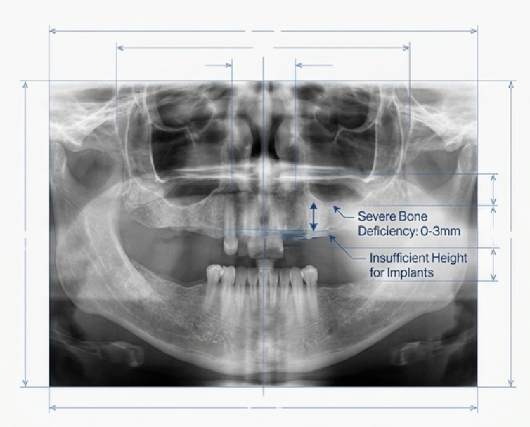
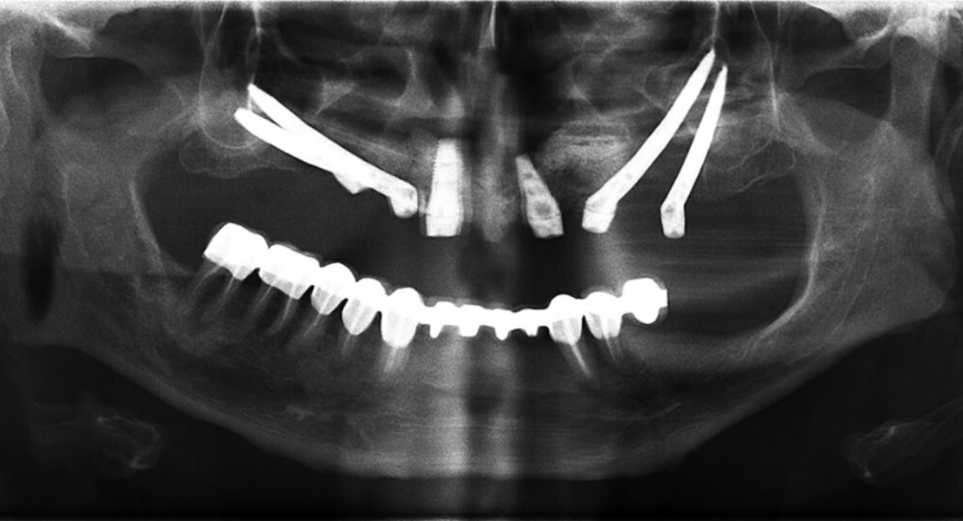
Implant Dentistry: X-rays may be used before dental implant installation to evaluate bone quality, determine implant appropriateness, and help plan surgery.
Oral Surgery: Prior to oral surgery procedures such as tooth extractions, impacted tooth removal, or jaw surgeries, multiple X-rays might be necessary to accurately diagnose the condition, plan the surgery, and ensure the safety and success of the procedure.
Full Mouth Reconstruction: Individuals undergoing extensive restorative treatments or full mouth reconstruction may require multiple X-rays within a short timeframe to assess the condition of the entire dentition, plan the treatment sequence, and ensure the desired outcomes.
Dental Emergencies and Trauma
In cases of dental emergencies or significant trauma to the oral structures, multiple X-rays within a month may be justified to assess the extent of the injury, guide emergency treatment decisions, and monitor the healing process.
Importance of Communication with Your Dentist
It’s important to keep lines of communication open with your dentist if dental X-rays are causing you anxiety. Dr. Chirag Chamria recommends that patients talk to their dentist about their concerns and queries regarding how often they need to be exposed to X-rays. Dentists may reassure patients regarding the safety of X-rays by explaining why they are necessary in each case.
How Dentists Minimise Radiation Exposure
Dental professionals should follow the principle of keeping radiation exposure as low as reasonably achievable while obtaining an image of adequate diagnostic quality. This can involve:
- Taking X-rays only when clinically justified
- Reviewing previous images before ordering new ones
- Selecting the smallest appropriate imaging area
- Using patient-specific exposure settings
- Using digital sensors and properly maintained equipment
- Avoiding repeat images caused by positioning or technical errors
- Selecting a limited-field CBCT instead of a larger scan when appropriate
Children require particular attention because developing tissues are more sensitive to radiation. Exposure settings and the imaging area should therefore be adjusted to the child’s size and diagnostic needs.
Should You Be Worried About Repeated Dental X-Rays?
Dental X-rays use ionising radiation, so exposure is not treated as completely risk-free. Nevertheless, the dose from properly performed conventional dental imaging is generally low. When an X-ray is clinically justified, the diagnostic benefit usually outweighs the small radiation-related risk. Patients should tell the dentist about recent dental or medical imaging and ask whether previous scans can be reviewed. This may prevent unnecessary duplication while ensuring that important dental disease is not missed.
Conclusion
In conclusion, the potential dangers of getting multiple dental X-rays in 30 days are relatively low. They emit minimal radiation, and dentists take precautions to minimize exposure. The frequency of X-rays depends on individual patient needs and is typically not monthly. While excessive radiation can pose risks, such instances are rare, and the benefits of diagnosing and treating dental issues often outweigh the minimal radiation exposure. It is essential for patients to communicate with their dentists and have a clear understanding of the reasons for X-ray recommendations. Ultimately, with proper safety measures and informed decision-making, dental X-rays remain an invaluable tool in ensuring oral health.
Frequently Asked Questions
Is it safe to have multiple dental X-rays in one day?
Manu small intraoral images may be safely taken during one appointment when they are required for diagnosis or treatment. The total exposure depends on the imaging technique and equipment—not merely the number of images.
Is it safe to have dental X-rays every month?
Monthly dental X-rays are not routinely required for most patients. However, they may occasionally be necessary during complex treatment, dental trauma, infection management or postoperative monitoring. Every additional image should have a clear clinical purpose.
Can dental X-rays cause cancer?
Dental X-rays involve low-dose ionising radiation, which carries a small theoretical risk. No imaging procedure involving ionising radiation should be performed unnecessarily. When properly selected and optimised, the clinical benefit of a required dental X-ray generally outweighs the low potential risk.
Are dental X-rays safe during pregnancy?
Necessary dental imaging can generally be performed during pregnancy using an appropriately selected and optimised technique. Patients should inform the dentist if they are pregnant or may be pregnant so that the urgency and imaging method can be assessed. Essential treatment for pain or infection should not be delayed without professional advice.
Are repeated dental X-rays safe for children?
Dental X-rays can be used safely in children when they are clinically necessary and the exposure settings are adjusted appropriately. Because children are more sensitive to radiation, the smallest suitable imaging area and child-specific settings should be used.
How can I know whether another dental X-ray is necessary?
Ask the dentist what the image is expected to show, how it will affect treatment and whether a previous X-ray can provide the required information. The decision should follow a clinical examination and consider the patient’s dental history and current symptoms.
Does radiation remain inside the body after a dental X-ray?
No. X-rays pass through the body during the exposure and do not remain inside it. However, radiation-related risk is considered across repeated exposures, which is why unnecessary imaging should still be avoided.
© All rights reserved by Royal Dental Implants Pvt Ltd
Issued in public interest