
Dental X-Rays | Radiovisiography v/s Orthopantomogram
What are Dental X-Rays?
Dental X-rays (radiographs) are the most common and widely available diagnostic imaging technique. Dental X-rays are pictures of the teeth, bones, and surrounding soft tissues to help find problems with teeth and bone.
What is Radiovisiography?
RVG is the latest X-ray technology in dentistry which helps in a better diagnosis. A high-quality digital image of root structures and bone can be obtained immediately. As compared to the conventional X-ray there is reduced radiation exposure to the patient.
What are Digital Radiography Dental X-Rays?
It is a novel method of medical imaging which uses digital X-ray sensors instead of conventional films to produce images. This digital RVG technology provides faster imaging, detailed radiographs and quality digital images. Digital radiography constitutes a system with components that include a digital image receptor, a digital image processing unit, an image management system, image and data storage devices, interface to a patient information system, a communication network and a display device with operated controls. Digital radiographs are stored in the form of digital data and can be displayed on a computer monitor.
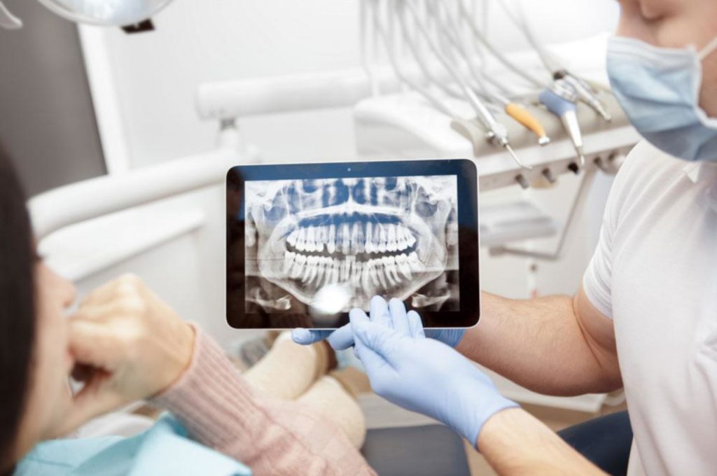
What is an Orthopantomogram (OPG)?
An Orthopantomogram (OPG) or panoramic dental X-ray can show a wide view of jaws, teeth and roots. It displays the nasal area, maxillary sinuses, jaw joints, teeth, upper jaw, lower jaw and surrounding bone on a single film. OPGs are used by Dentists to determine the status of wisdom teeth, to reveal cysts, tumours, bone irregularities and more.
Two types of x-rays are used in dentistry
- Intraoral – IOPA, RVG
- extraoral – OPG, CBCT
How does Dental RVG X-Rays work?
RVG means Radio Visio Grapghy. It is an instant method of taking dental x-rays. It is a form of intraoral digital imaging which uses advanced sensors instead of traditional films. The image captures better details and also gives lesser radiation as compared to traditional image techniques. There is around 80% reduction in dosage as compared with conventional x-rays. It provides quick results and aids in early diagnosis without wasting patients time.
It is localized and limited to one or 3 to 4 teeth. This can be used as a chair side tool during treatment. In periodontal diseases, it helps to evaluate the alveolar bone loss. RVG is used to educate and motivate the patient. RVG takes localised area images, hence multiple dental implant x-rays are not possible and also as it causes contamination of the surgical area. Multiple RVGs cause patient’s discomfort and it is time consuming where one-day dental implants are choice of treatment. An effective dose is 1-8 micro SV.

How does Dental OPG X-Ray work?
ortho Panto Gram is done for upper and lower teeth with jawbones and maxillary, nasal sinus. Generally recommended for full mouth cases. OPG is commonly used to diagnose teeth impaction, fractures, dislocation, infection, tumours, sinuses. Recommended in patients with severe gag during placement of RVG sensor. It can be used for diagnostic purpose. For dental implant planning, OPG is advisable compared to RVG as the entire bone structure, nerve pathway can be assessed and implant placements can be done accordingly. Effective doses are 4-30 micro SV.
Leave a reply
Leave a reply