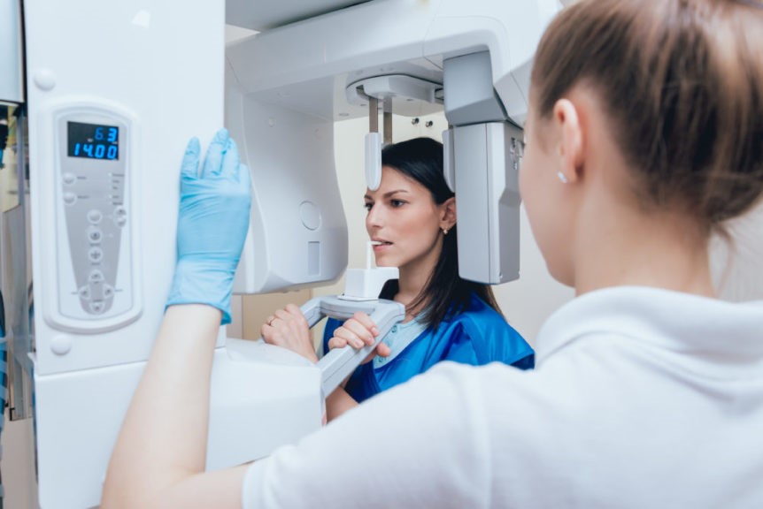
Dental caries, commonly known as tooth decay, is a bacterial infections of the teeth. When left untreated, it can lead to further complications and even tooth loss. The prevalence of dental caries has been increasing in recent years. According to a study by the Centers for Disease Control and Prevention, some 20% of all adults in the U.S. have untreated cavities, making it one of the most frequent oral diseases today. These facts should motivate dentists to adopt new strategies for fighting tooth decay and identifying individuals who are at risk of developing it as early as possible. CBCT technology provides just such an opportunity. Here is everything you need to know about cross-sectional imaging and whether or not your dentist can assess tooth decay!
What is cross-sectional or CBCT imaging?
Cross-sectional imaging is a non-invasive imaging technique that allows researchers to study teeth and other dental structures in cross-sections. To do so, they will generally use a technique called ultrasound biomicroscopy. This technique relies on the generation of ultrasonic waves, which then reflect on the object of interest. The frequency of the reflected waves reveals the object’s physical properties, such as density, thickness, and shape.

These waves are also detectable by our bodies, and as such, they can be used as a diagnostic tool. In dentistry, they have been applied to the imaging of tooth surfaces and interiors. The results obtained using this technique are comparable to the outcomes of conventional X-ray imaging.
Just like with conventional X-ray imaging, cross-sectional imaging relies on the passing of electromagnetic waves through the object of interest. However, for cross-sectional imaging, only a single wavelength of electromagnetic waves is used. These waves are reflected by the object and then collected by a detector placed opposite the source. The detector then transforms the reflected waves into an image.
For teeth and other dental structures, the electromagnetic waves used for cross-sectional imaging are in the ultrasound frequency range. The technical name for this imaging modality is ultrasound biomicroscopy.
What can dentist learn from CBCT imaging?
Besides being an excellent diagnostic tool for dental structures, cross-sectional imaging also provides non-invasive access to the oral cavity, which makes it particularly suitable for children. That being said, there is a lot that can be learned from cross-sectional imaging. For example, dentists can visualize the tooth’s surface, its underlying structures (e.g. supporting tissues, nerves, and blood vessels), cavities, tooth defects, and other pathological changes. Additionally, they can use the technique to chart the tooth’s growth, which can help with prognostication and treatment planning.
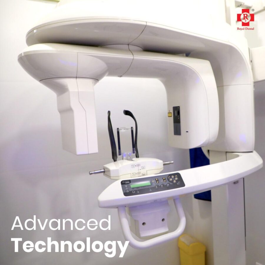
Cross-sectional imaging can also be used to study the tooth’s microstructure, which is important for developing new preventive and restorative dental treatments. For example, the technique has been used to develop new tooth-colored fillings and sealants. While the aforementioned uses of cross-sectional imaging are important and can be of great help to dentists, they are not directly related to the detection of tooth decay.
But, as described in the introduction, dental caries is a bacterial infection of the teeth and, as such, it leaves some traces. So, while there is no direct way to directly assess tooth decay via cross-sectional imaging, there are ways to indirectly detect it. These rely on the presence of certain signs that are indicative of tooth decay.
Uses of CBCT for tooth infection
Dental CBCT systems have been sold in the United States since the early 2000s and are increasingly used by radiologists and dental professionals for various clinical applications including dental implant planning, visualization of abnormal teeth, evaluation of the jaws and face, cleft palate assessment, diagnosis of dental caries (cavities), endodontic (root canal) diagnosis, and diagnosis of dental trauma.

For example, the presence of a dip in the tooth surface (indicating a carious lesion) or a thickening of the tooth structure (indicating dental calculus) can be detected via cross-sectional imaging. The image processing techniques used for image analysis can also be used to quantify the amount of bacterial growth on the tooth surface. This can be used as a proxy for dental caries, as a high bacterial growth rate is indicative of dental caries.
Conclusion
Cross-sectional imaging is a promising imaging modality that has been demonstrated to be effective for dental imaging. It allows dentists to visualise the tooth’s surface and underlying structures, including the bacterial growth on the tooth surface. This is important for developing new preventive and restorative dental treatments. Moreover, cross-sectional imaging can be used to study the tooth’s microstructure. Which is important for developing new tooth-colored fillings and sealants.
While cross-sectional imaging cannot be used directly to assess tooth infection. Its indirect detection can be used to assess tooth decay. Besides being an excellent diagnostic tool for dental structures. Cross-sectional imaging also provides non-invasive access to the oral cavity, which makes it particularly suitable for children.

